

Thrombosis Models
Leveraging a well-established in vivo pharmacology platform and an experienced scientific team, Greentech provides comprehensive dementia animal models and standardized in vivo efficacy evaluation services for global clients. Our solutions support disease mechanism studies, target validation, and the nonclinical development of therapeutics for neurodegenerative disorders.
Our Dementia Models
1. APP/PS1 Double Transgenic Mouse Model
APP/PS1 double transgenic mice are a well-established Alzheimer's disease (AD) model that develops progressive Aβ deposition, amyloid plaque formation, neuronal degeneration, and cognitive impairment. Amyloid plaques emerge at approximately 4 months of age, followed by robust Aβ accumulation by 6 months and significant learning and memory deficits between 10 and 12 months.
Applications: Since APP/PS1 mice do not develop neurofibrillary tangles (NFTs), they are well suited for the nonclinical evaluation of Aβ-targeted therapeutics.
Limitations: Lack of prominent neurodegeneration, brain atrophy, and NFT formation.
2. 5×FAD Mouse Model
The 5×FAD mouse is a well-established Alzheimer's disease (AD) model carrying five familial AD-associated mutations in the human APP and PSEN1 genes. The model exhibits early and robust amyloid pathology, neuroinflammation, synaptic loss, and neuronal degeneration, making it one of the most widely used models for preclinical AD research.
Applications: Suitable for investigating the pathological mechanisms of Alzheimer's disease, the role of immune responses in neurodegeneration, and the preclinical development of Alzheimer's disease therapeutics.
Limitations: Does not develop neurofibrillary tangles (NFTs).
3. SAMP8 Mouse Model – Accelerated Senescence Model
SAMP8 mice are a well-established aging-associated Alzheimer's disease (AD) model characterized by accelerated aging, progressive cognitive decline, increased APP expression, Aβ- and tau-related pathology, oxidative stress, and cholinergic dysfunction, closely resembling key pathological features of human AD.
Applications: Since APP/PS1 mice do not develop neurofibrillary tangles (NFTs), they are well suited for the nonclinical evaluation of Aβ-targeted therapeutics.
Limitations: Lack of prominent neurodegeneration, brain atrophy, and NFT formation.
4. Diabetes-Associated Cognitive Impairment Models
Diabetes-associated cognitive impairment involves impaired hippocampal synaptic plasticity, reduced neurotrophic factor levels, increased blood–brain barrier permeability, neuroinflammation, and neuronal apoptosis.
Models:
1) db/db mouse model
2) High-fat diet (HFD) combined with streptozotocin (STZ)-induced diabetic rat model
5. 2-Vessel Occlusion (2-VO) Vascular Dementia Model
Vascular dementia (VD) results from impaired cerebral blood flow and is characterized by cognitive dysfunction. The two-vessel occlusion (2-VO) model induces chronic cerebral hypoperfusion and secondary white matter injury and is widely used in vascular dementia research and drug evaluation. The model is established by ligation of the bilateral common carotid arteries and is relatively simple, reproducible, and cost-effective.
Applications: Suitable for investigating the pathogenesis and pathological changes of vascular dementia, cognitive impairment, and for pharmacodynamic screening and evaluation.
6. Scopolamine-Induced Model – Central Cholinergic System Impairment
Scopolamine crosses the blood–brain barrier and disrupts the central cholinergic system, causing short-term learning and memory impairment. Scopolamine administration can reduce brain acetylcholine (ACh) levels and induce Alzheimer's disease-like cognitive deficits. A single administration can produce significant learning and memory impairment within approximately 30 minutes, while repeated administration for 4 weeks increases Aβ and hyperphosphorylated Tau levels in the rat brain.
Applications: Suitable for investigating cognitive impairment in Alzheimer's disease models and evaluating related drugs.
Limitations:
1) Dementia-like symptoms are reversible.
2) Non-specific blockade of central muscarinic (M) receptors may cause behavioral effects such as anxiety, reduced locomotor activity, and increased spontaneous activity, which may interfere with behavioral assessments.
Study Endpoints
1) Neurobehavioral assessments:Morris water maze (MWM), including acquisition and probe trials; passive avoidance test; novel object recognition test; Barnes maze; social interaction test; open field test; rotarod test; elevated plus maze; and other behavioral assessments.
2) Aβ detection:
a. ELISA-based quantification of Aβ levels in tissue homogenates, plasma, and cerebrospinal fluid (CSF) b. Immunohistochemical staining of amyloid plaques
3) Tau protein detection: Western blot (WB) analysis of hyperphosphorylated Tau levels in the brain.
4) Histopathological and immunohistochemical assessments:
a. Neuronal number
b. Microglial cell number
c. Astrocyte number
d. Neurofibrillary tangles (NFTs)
5) Cholinergic system assessment: Measurement of AChE and ChAT activities and ACh levels in the hippocampus and cortex by ELISA, particularly for scopolamine-induced models.
6) Neuroinflammation assessment
7) Oxidative stress assessment
Representative Study
1. APP/PS1 Double Transgenic Mouse Model of Alzheimer's Disease
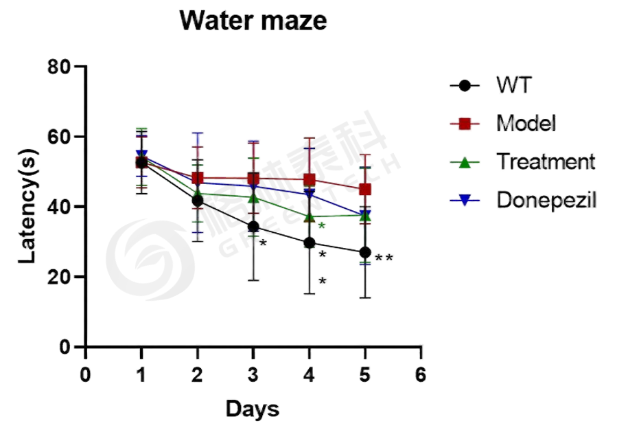
Figure 1. Morris water maze assessment in the APP/PS1 double transgenic mouse model.
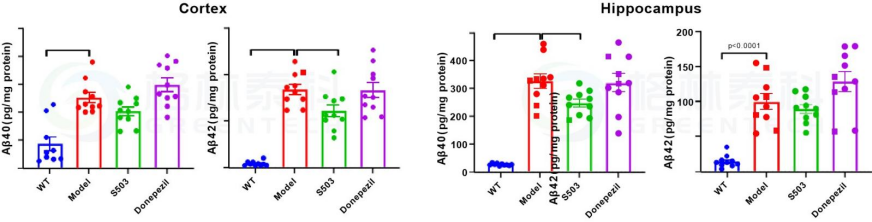
Figure 2. Aβ40 and Aβ42 levels in the cerebral cortex and hippocampus of APP/PS1 mice.
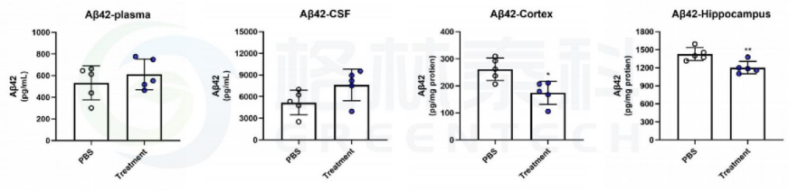
Figure 3. Aβ42 levels in plasma, cerebrospinal fluid, cortex, and hippocampus of APP/PS1 mice.
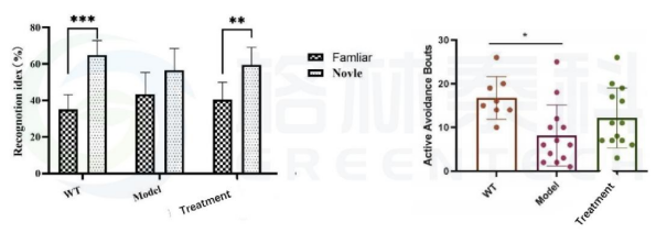
Figure 4. Behavioral assessments in APP/PS1 mice: novel object recognition and shuttle box tests.

Figure 5. Histopathological assessment of APP/PS1 mouse brain tissue: Aβ and GFAP staining.
2. Neurofunctional Decline in 5xFAD Mice
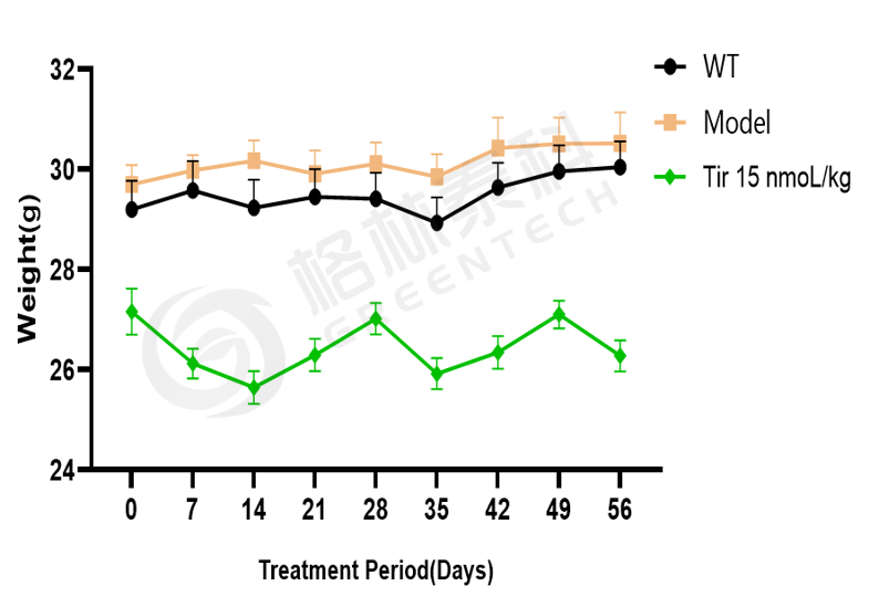
Figure 6. Body weight changes in 5xFAD mice.
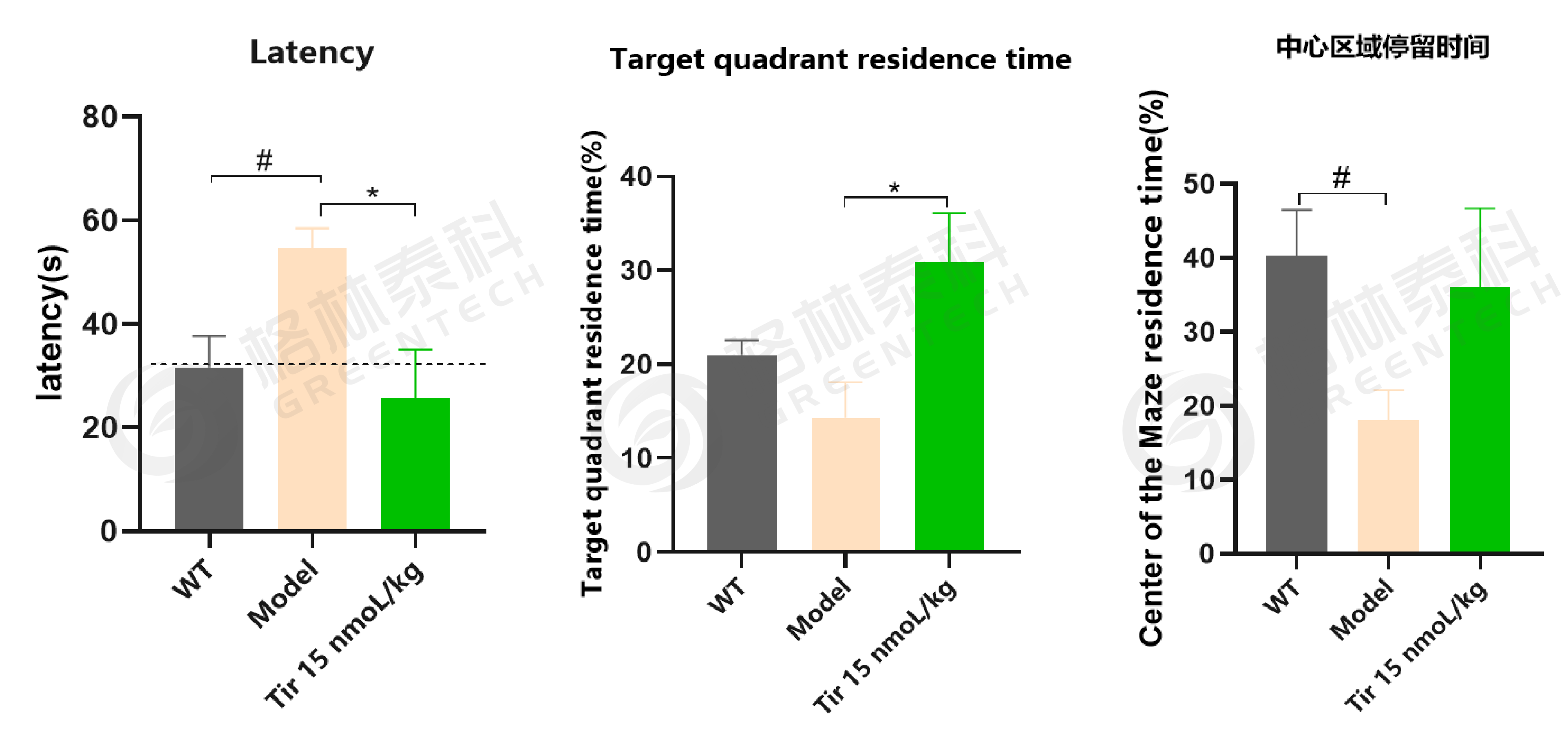
Figure 7. Morris water maze results in the 5xFAD mouse model after 9 weeks of treatment.
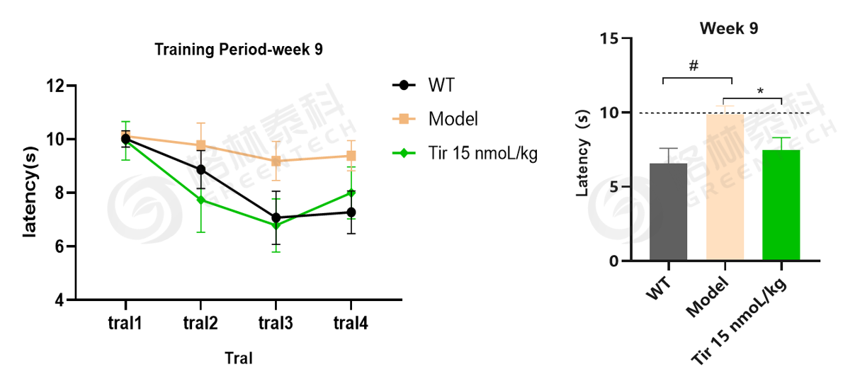
Figure 8. Shuttle box test results in the 5xFAD mouse model after 9 weeks of treatment.
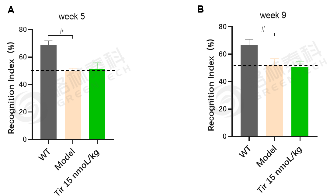
Figure 9. Novel object recognition test results in the 5xFAD mouse model.
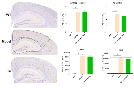
Figure 10. Aβ staining results in brain tissue from the 5xFAD mouse model.
Inquiries
Request a quote now, or email us at BD@greentech-bio.com to inquire about our services or obtain a quote for your project.
References
1. Congdon EE, Sigurdsson EM. Tau-targeting therapies for Alzheimer disease. Nature Reviews Neurology. 2018;14(7):399–415.
2. Götz J, Bodea LG, Goedert M. Rodent models for Alzheimer disease. Nature Reviews Neuroscience. 2018;19(10):583–598.
3. Canudas AM, Gutierrez-Cuesta J, Rodríguez MI, et al. Hyperphosphorylation of microtubule-associated protein tau in senescence-accelerated mouse (SAM). Mechanisms of Ageing and Development. 2005;126(12):1300–1304.
4. Porquet D, Andrés-Benito P, Griñán-Ferré C, et al. Amyloid and tau pathology of familial Alzheimer's disease APP/PS1 mouse model in a senescence phenotype background (SAMP8). Age. 2015;37(1):9747.
5. Das TK, Jana P, Chakrabarti SK, Abdul Hamid MRW. Curcumin downregulates GSK3 and Cdk5 in scopolamine-induced Alzheimer's disease rats, abrogating Aβ40/42 and tau hyperphosphorylation. Journal of Alzheimer's Disease Reports. 2019;3(1):257–267.
6. Huang LJ, Zhao CY, Feng XH, et al. Exploration of a nonclinical pharmacodynamic evaluation system for anti-Alzheimer's disease drugs. Acta Pharmaceutica Sinica. 2020;55(5):35–51.
7. Forner S, et al. Systematic phenotyping and characterization of the 5xFAD mouse model of Alzheimer's disease. Scientific Data. 2021;8(1):270.
8. Ruan S, et al. Aerobic exercise alleviates cognitive impairment in T2DM mice through gut microbiota. Scientific Reports. 2025;15(1):23917.
-
- CRO Services
- Cardiovascular Diseases
- Psychiatry & Neurology
- Oncology
- Metabolic Diseases
- Inflammation & Autoimmune Diseases
- Pain & Analgesia
- Bone, Joint & Cartilage Diseases
- DMPK & Non-GLP Toxicology
- In Vitro Drug Screening
- Efficacy Testing for Medical Devices
-
- Platforms
- Endovascular Surgery
- Behavioral Tests
- Histopathology
- Clinical Testing
- Bioanalysis
-
- Resources
- Brochure
- FAQ
-
- About Us
- Company Profile
- Company Culture
- Honors
- Cooperators
- Enterprise Memorabilia
-
- News & Blog
- Company News
- Blog
-
- Contact Us