Models of Ischemic Stroke
Greentech Bioscience offers well-characterized ischemic stroke models to help clients study ischemic stroke injury and mechanisms and assess effects of therapies on ischemic stroke. Our stroke models have proven reproducible, reliable, and effective in a number of studies, including middle cerebral artery occlusion (MCAO) models in rodents and non-human primates, as well as photothrombotic ischemia model.
Our Animal Models of Ischemic Stroke
1. Middle Cerebral Artery Occlusion (MCAO) Model in Rodents
Induction: Occlusion of the middle cerebral artery (MCA) occlusion by a suture
Species: Rats
Disease characteristics:
- Well reproduce clinical MCA infarcts
- Exhibiting a well-defined ischemic penumbra (hypoperfused tissue at risk of infarction but can be potentially salvaged)
- Reproducible deficits in sensorimotor and cognition
- Can be used to evaluate neuroprotective drugs
- Not suitable for thrombolytic drug research
- A long ischemia time leading to greater mortality risk
- Classified into ischemia-reperfusion model and permanent ischemia model
The Difference Between Reperfusion Model and Permanent Ischemia Model
- Ischemia-reperfusion models consist of primary brain tissue injury and secondary damage caused by energy metabolism disorders after hypoxic reperfusion, with a time difference between the two injuries; The permanent ischemia model does not have this feature.
- The infarction core in the permanent infarction model is larger than that of the ischemic reperfusion model, and the ischemic penumbra may be smaller.
- The mortality rate of the permanent infarction model is relatively higher.
Clinical Assessment
- Behavioral tests
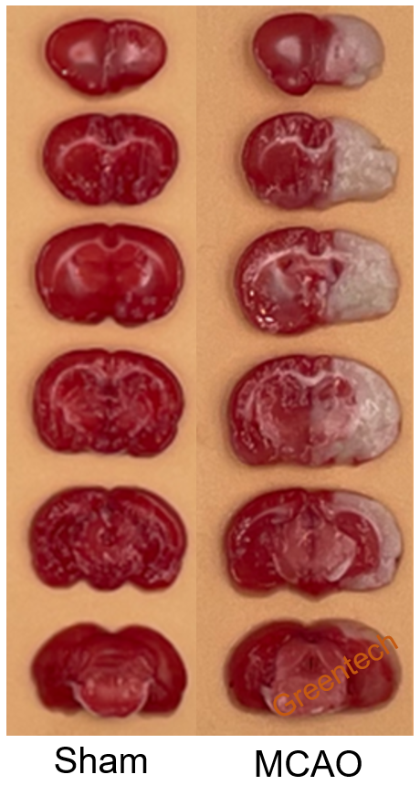
- Infarct volume (TTC staining)
- Grid test
- Novel object recognition test
Basic Service
- Body weight
- Blood and tissue collection
- Histopathology
- Cerebral blood flow
Case Study
Animal model of stroke model

Figure 1. TTC staining of cerebral tissue in the MCAO rat model
2. MCAO model in non-human primates (NHPs)
Induction: Coil embolization of the right middle cerebral artery (MCA) based on digital subtraction angiography (DSA)
Species: rhesus monkeys
Disease characteristics:
MCAO model in NHPs can reproduce the clinical infarct size (generally 1.67%~6.67%), while MCAO rodents exhibit much larger infarct size (11%~24%). Moreover, NHPs and human have similar cerebrovascular anatomy, neurological function and immunological characteristics. Consequently, NHPs are considered as optimal model for assessing anti-stroke drugs and endovascular treatment.
Clinical Assessment
- Neurological deficit score
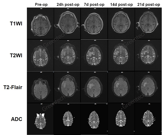
- Infarct volume (MRI, multiple scanning sequences available)
Basic Service
- Body weight
- Blood and tissue collection
- Histopathology
- Cerebral blood flow
Case Study
Rhesus monkey model of ischemic stroke

Figure2. DSA showing the occlusion and reperfusion of the right middle cerebral artery of a Rhesus monkey.

Figure 3. MRI scanning pre and post surgery. MRI scanning sequences: T1WI, T1 weighted image; T2WI, T2 weighted image; T2-Flair, T2-weighted-Fluid-Attenuated Inversion Recovery; ADC, apparent diffusion coefficient.
3. Photothrombotic Ischemia Model in Rodents
Induction: Photo-activation of a previously injected light-sensitive dye to induce an ischemic damage within a given cortical area
Species: rats
Disease Characteristics: The photothrombotic stroke model results in smaller infarct volumes than MCAO rodent models. Photothrombotic stroke model is featured by low invasion, high reproducibility, and ischemic lesion location.
Clinical Assessment
- Behavioral tests
- Infarct volume (TTC staining)
- Grid test
- Novel object recognition test
Basic Service
- Body weight
- Blood and tissue collection
- Histopathology
- Cerebral blood flow
References
1. Kito G , Nishimura A , Susumu T , Nagata R , Kuge Y , Yokota C , Minematsu K. Experimental thromboembolic stroke in cynomolgus monkey.Journal of Neuroscience Methods 105 (2001) 45–53.
2. Makoto Sasaki , Kohsuke Kudo, Kaneyoshi Honjo, Jin-Qing Hu, Hai-Bin Wang, Katsuya Shintaku. Prediction of infarct volume and neurologic outcome by using automated multiparametric perfusion-weighted magnetic resonance imaging in a primate model of permanent middle cerebral artery occlusion. Journal of Cerebral Blood Flow & Metabolism (2011) 31, 448–456.
3. Sommer Clemens J. Ischemic stroke: experimental models and reality. Acta Neuropathol (2017) 133:245–261.
Inquiries
Request a quote now, or email us at BD@greentech-bio.com to inquire about our services or obtain a quote for your project.


