Models of Liver Injury, Cirrhosis, Failure
Greentech provides a broad spectrum of animal models of liver diseases for drug efficacy testing including animal models of acute hepatic damage, animal models of liver cirrhosis, and animal models of liver failure. We have extensive experience in preclinical pharmacology studies, especially metabolic diseases, and are capable of customizing animal models according to clients’ needs.
Our Animal Models of Liver Damage, Cirrhosis, Failure
Models | Species | Induction | Induction Period |
CCl4 induced acute liver injury model | Mice, rats | Intraperitoneal injection of CCl4 | - |
Acute alcohol induced acute liver injury model | Mice, rats | Oral gavage of ethanol solution for three times | - |
Acetaminophen induced acute liver injury model | Mice, rats | Intraperitoneal injection of acetaminophen | - |
D-gal induced acute liver failure model | Mice, rats | Intraperitoneal injection of D-gal | - |
ANIT induced primary biliary cirrhosis (PBC) model | SD rats, Wistar rats, C57BL/6J mice | Oral gavage of ANIT | 4 weeks |
Bile duct ligation (BDL) induced PBC model | SD rats, Wistar rats | Bile duct ligation | 2~3 weeks |
2-OA/BSA induced PBC model | C57BL/6J mice | Intraperitoneal injection of the combination of 2-OA and BSA | 12~16 weeks |
DDC induced primary sclerosing cholangitis (PSC) model | C57BL/6J mice | 3,5-diethoxycarbonyl-1,4-dihydrocollidine (DDC) diet | 8 weeks |
BDL induced acute liver failure model | Rhesus monkeys/cynomolgus monkeys | Bile duct ligation | Fibrosis occurs at 4 week post surgery; cirrhosis appears at 8 week post surgery. |
Clinical Assessment
(1) Body weight, liver index, food intake
(2) Liver function: ALT, AST, ALB, LDH, etc.
(3) Lipid metabolism: TC, HDL, LDL, TG, etc.
(4) Cholestasis: T-BIL, D-BIL, I-BIL
(5) Biliary obstruction: ALP, GGT
(6) Hepatic histopathology: HE, MASSON staining
(7) Hepatic fibrosis: detection of collagen and hydroxyproline
(8) Imaging: color ultrasound, CT, PET/CT, etc.
Case Study
1. BDL induced SD rat PBC models
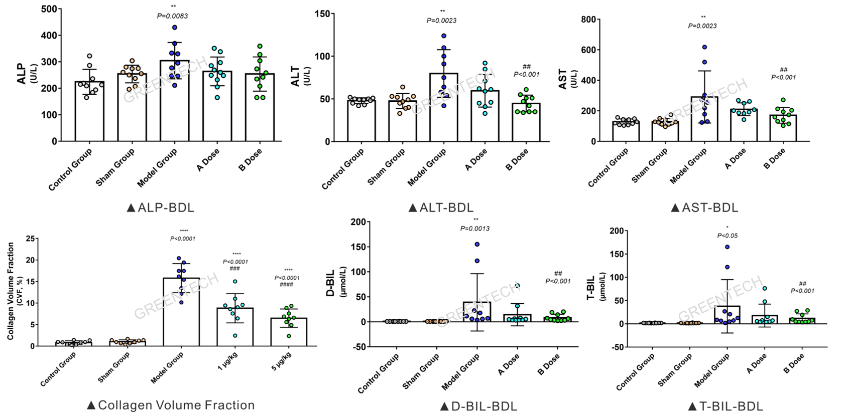
Figure 1. Evaluation of function of liver and gall, hepatic collagen volume fraction 2 weeks after bile duct ligation in SD rats.

Figure 2. Hepatic histopathology (HE/SR staining, immunohistochemical staining of α-SMA) 2 weeks after bile duct ligation in SD rats.
2. ANIT induced rat PBC models


Figure 3. Evaluation of function of liver and gall in SD rats 4 weeks after ANIT induction.
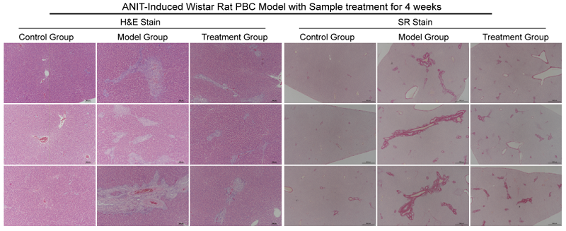
Figure 4. Hepatic histopathology (HE and SR staining) of SD rats 4 weeks after ANIT induction.
3. DDC induced mouse PBC models

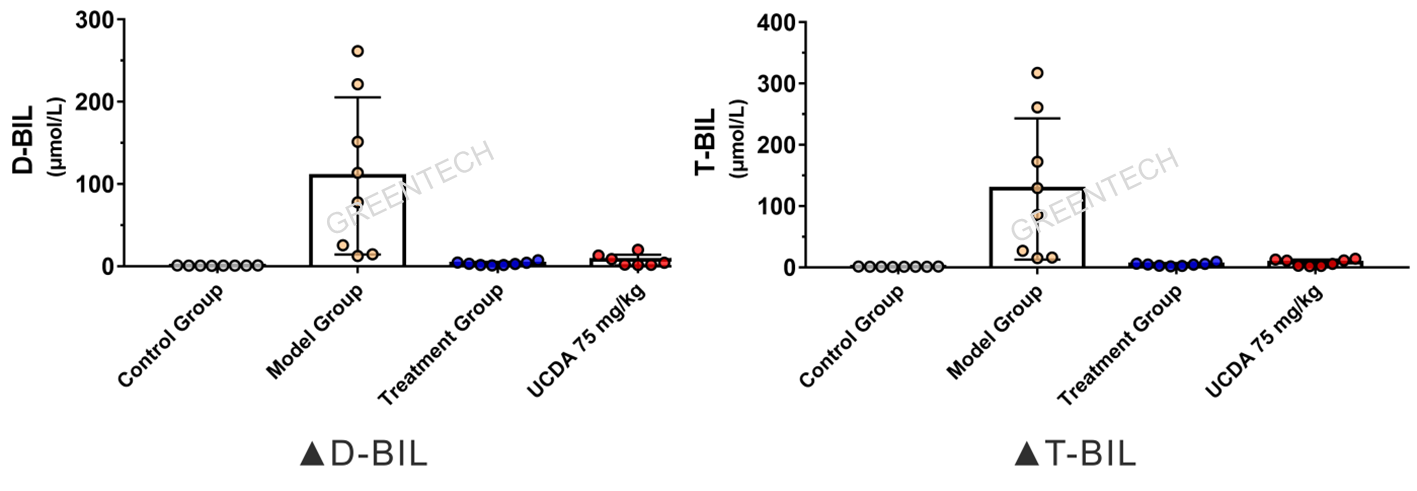
Figure 5. Evaluation of function of liver and gall in C57 mice feeding on 0.1% DDC diet for 8 weeks.
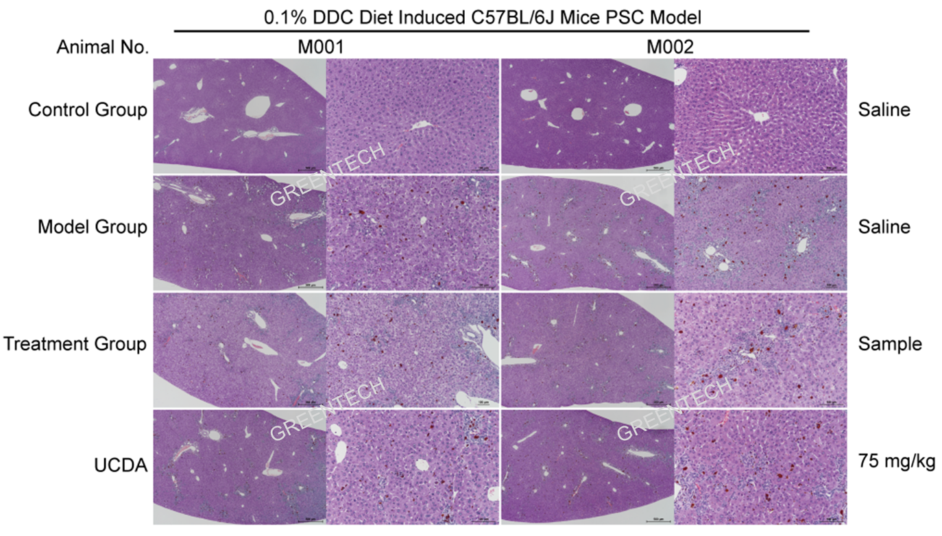
Figure 6. Hepatic histopathology (HE staining) of C57 mice feeding on 0.1% DDC diet for 8 weeks.
4. BDL induced NHP models of liver fibrosis, cirrhosis, and failure

Figure 7. Blood biochemistry analysis of cynomolgus monkeys 8 weeks after BDL.
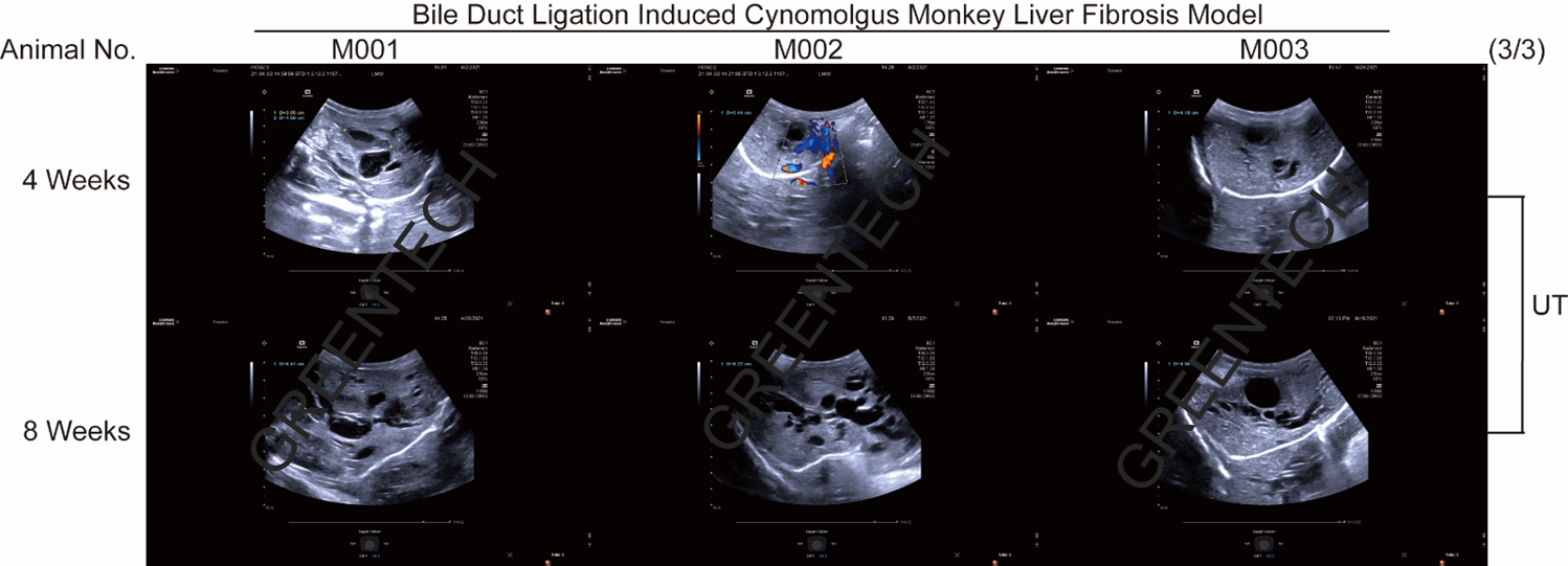
Figure 8. Ultrasound examination of liver fibrosis in cynomolgus monkeys after BDL.
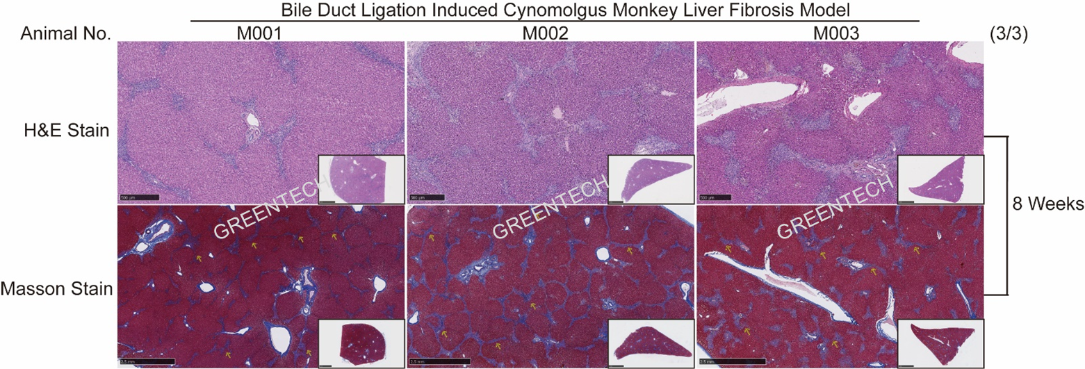
Figure 9. H&E and Masson staining demonstrated distinct cholestasis-induced model of liver fibrosis/cirrhosis in cynomolgus monkeys 8 weeks after BDL.
References
1. Dongying W, et al. Protective effects of Ziyang tea polysaccharides on CCl4-induced oxidative liver damage in mice. 2014;143:371-378.
2. Coccolini F, Coimbra R, Ordonez C, et al. Liver trauma: WSES 2020 guidelines[J]. World Journal of Emergency Surgery, 2020, 15: 1-15.
3. Fujiwara K, et al. Cite Share Intravascular coagulation in acute liver failure in rats and its treatment with antithrombin III. Gut. 1988;29(8):1103-8.
4. Julian A Luetkens, et al. Quantification of Liver Fibrosis at T1 and T2 Mapping with Extracellular Volume Fraction MRI: Preclinical Results. Radiology. 2018;288(3):748-754.
Inquiries
Request a quote now, or email us at BD@greentech-bio.com to inquire about our services or obtain a quote for your project.


