

Pulmonary Embolism (PE) Models
Greentech provides validated autologous thrombus-induced pulmonary embolism (PE) models in rats, mice, dogs, and non-human primates (NHPs), together with comprehensive in vivo pharmacology and pharmacodynamic services to support the discovery and preclinical development of antithrombotic and thrombolytic therapies. Our studies are designed to support both domestic and international IND programs.
Our Pulmonary Embolism (PE) Models
Greentech has established a sodium laurate-induced rat TAO model through femoral artery injection. This well-characterized model reproduces key pathological features of thromboangiitis obliterans and is widely used for preclinical efficacy evaluation of candidate therapeutics.
Animals: Rat, Mouse, Dog, Non-human primate (NHP)
Induction: Autologous blood is collected from each animal and processed into thrombi of predefined size before embolization:
1) Rat: Cylindrical autologous thrombi are prepared and injected via the right jugular vein.
2) Mouse: Autologous thrombus suspension is prepared and administered through the jugular vein.
3) Dog: Cylindrical autologous thrombi are delivered into the main pulmonary artery using a minimally invasive interventional procedure.
4) NHP: Customized protocols are available upon request.
Characteristics: The autologous thrombus-induced PE model produces rapid and reproducible pulmonary embolism, making it well suited for the preclinical evaluation of thrombolytic agents and other investigational therapies.
Positive Control Drugs: commonly used positive controls include: recombinant human tenecteplase (rh TNK-tPA) / recombinant tissue plasminogen activator (rt-PA)
Study Endpoints
Greentech offers comprehensive efficacy assessment, including
1) Coagulation parameters (e.g., fibrinogen [FIB], thrombin time [TT])
2) Histopathological quantification of pulmonary thrombi
3) Pulmonary angiography (dog and NHP models)
4) Optional endpoints, including hemodynamic assessment and plasma biomarker analysis
Representative Studies
1. Mouse Autologous Thrombus-Induced Pulmonary Embolism Model
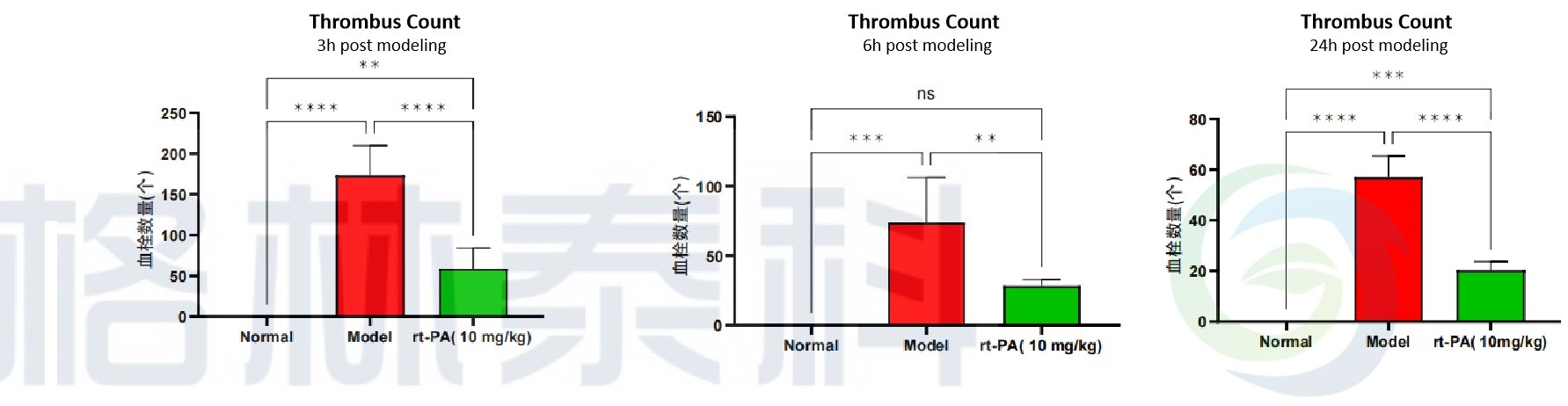
Figure 1. Histopathological quantification of pulmonary thrombi in the mouse autologous thrombus-induced pulmonary embolism model.
2. Dog Autologous Thrombus-Induced Pulmonary Embolism Model
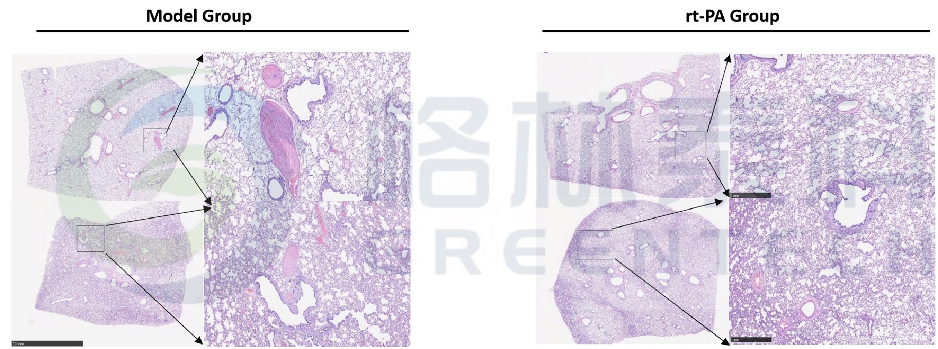
Figure 2. Representative histopathological findings in the dog autologous thrombus-induced pulmonary embolism model. Extensive thrombi were observed in pulmonary arteries of the model control group, whereas only small residual thrombi were detected in animals treated with rt-PA.
References
Zheng R, Liu J, Wan J, et al. Establishment and comparison of acute pulmonary thromboembolism models in mice. Chongqing Medicine. 2012;41(5):428–431.
Feng W, Zhang Y, Sun F, et al. Establishment of an autologous pulmonary thromboembolism model in rats. Journal of Clinical Pulmonary Medicine. 2015;(5):785–788.
Inquiries
Request a quote now, or email us at BD@greentech-bio.com to inquire about our services or obtain a quote for your project.
-
- CRO Services
- Cardiovascular Diseases
- Psychiatry & Neurology
- Oncology
- Metabolic Diseases
- Inflammation & Autoimmune Diseases
- Pain & Analgesia
- Bone, Joint & Cartilage Diseases
- DMPK & Non-GLP Toxicology
- In Vitro Drug Screening
- Efficacy Testing for Medical Devices
-
- Platforms
- Endovascular Surgery
- Behavioral Tests
- Histopathology
- Clinical Testing
- Bioanalysis
-
- Resources
- Brochure
- FAQ
-
- About Us
- Company Profile
- Company Culture
- Honors
- Cooperators
- Enterprise Memorabilia
-
- News & Blog
- Company News
- Blog
-
- Contact Us