Models of Hyperuricemia & Gout
Greentech provides validated hyperuricemia model, MSU air pouch model, and MSU-induced gouty arthritis model for compound testing. We have extensive experience in preclinical pharmacology studies, and are capable of customizing animal models according to clients’ needs.
Our Animal Models of Hyperuricemia and Gout
1. Animal model of hyperuricemia
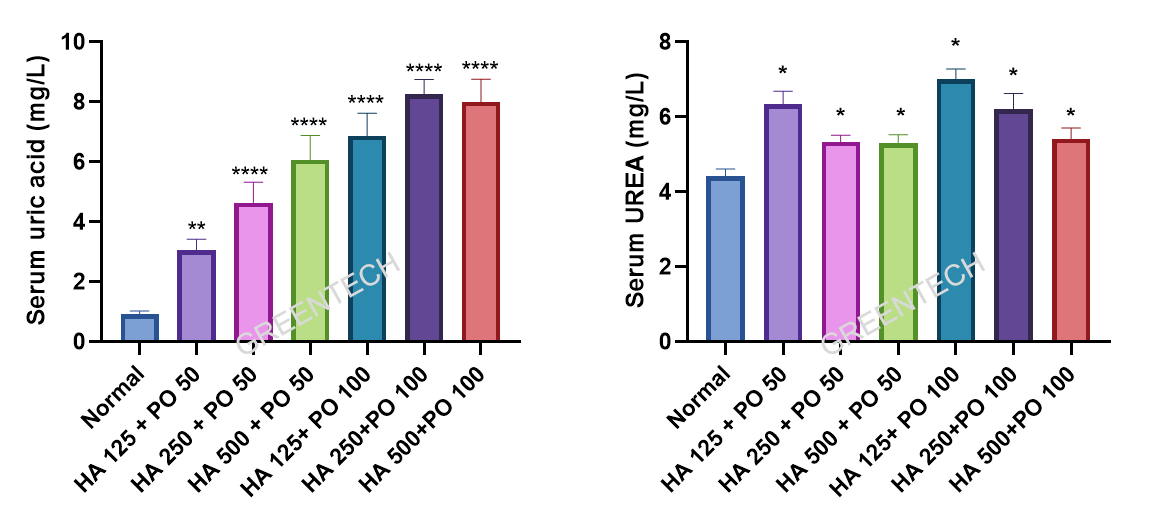
At present, there is no stable, simple, or robust animal models of hyperuricemia. Combination of multiple factors such as increase of uric acid precursors and inhibition of uricase activity and uric acid excretion, can induce hyperuricemia in rodents such as rats to some extent. We provide the model of hyperuricemia induced by the combination of hypoxanthine (HX) and potassium oxonate (PO). Hypoxanthine is an intermediate metabolite of the purine metabolism, and potassium oxonate is a uricase inhibitor. Oral gavage of PO and HX is most commonly used to induce hyperuricemia, which is reported in various literature.
Animal Species
Mice, rats
Clinical Assessment
Serum uric acid (UA), serum creatinine (Cr), serum urea nitrogen (BUN)
Case Study
Rat model of hyperuricemia

Figure 1. The serum UA and UREA levels of SD rat hyperuricemia model with different doses of hypoxanthine and potassium oxonate.
2 Models of Gout
2.1 MSU Air Pouch Model
Greentech Bioscience provides gout air pouch model which accurately mimics human gout. This acute uratic inflammation mouse model is induced by subcutaneous injections of monosodium urate (MSU) crystals in mice. In the MSU air pouch model, quantification of inflammatory response and histological examination of the pouch lining provide evidence of a protective effect of a test compound.
2.2 MSU Induced Gouty Arthritis Model
Acute gouty arthritis model is set up by given intra-articular injection of MSU crystal suspension into one knee joint. This gouty arthritis model provides a good, simple screening tool to test anti-gouty arthritis compounds.
Animal Species
Mice, rats, rabbits
Clinical Assessment
(1) Clinical observations and body weight measurement
(2) Caliper measurements of joint or air pouch swelling
(3) Pain assessment and joint dysfunction score
(4) Inflammation evaluation: WBC, IL-1β, IL-6, IL-8, TNF-α, PGE2
(5) Histopathological assessment
Case Study
1. MSU air pouch model
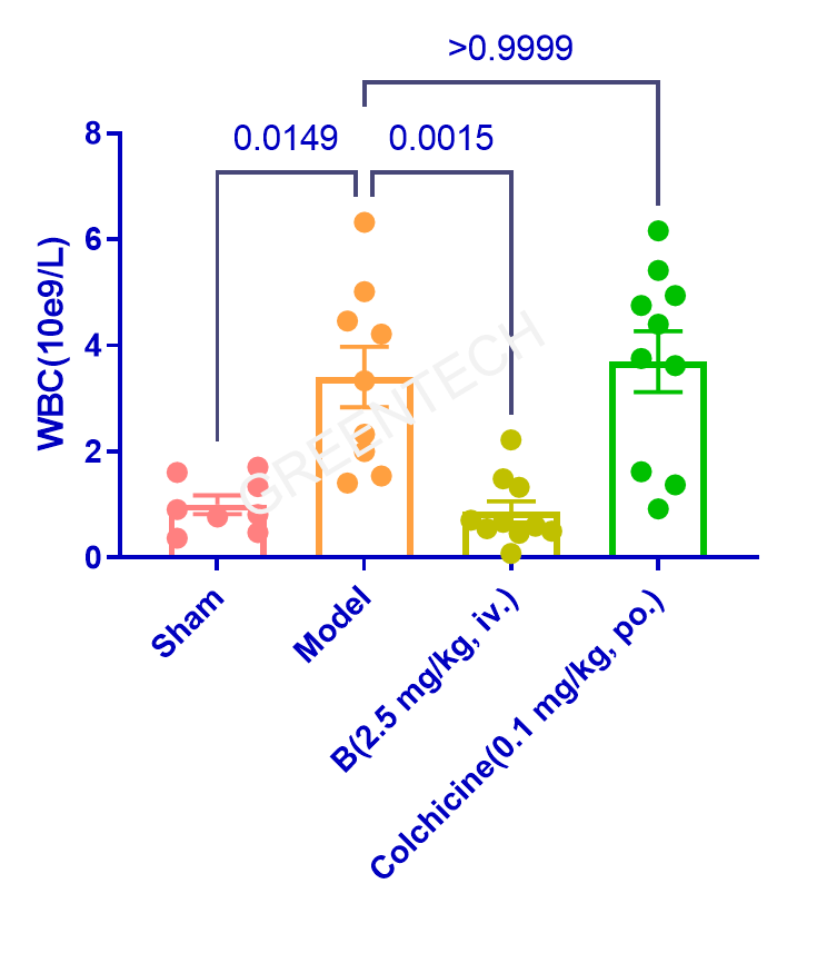
Figure 2. White blood cell (WBC) count of air pouch exudate in SD rats.
2. Rabbit model of MSU crystal-induced acute gouty arthritis

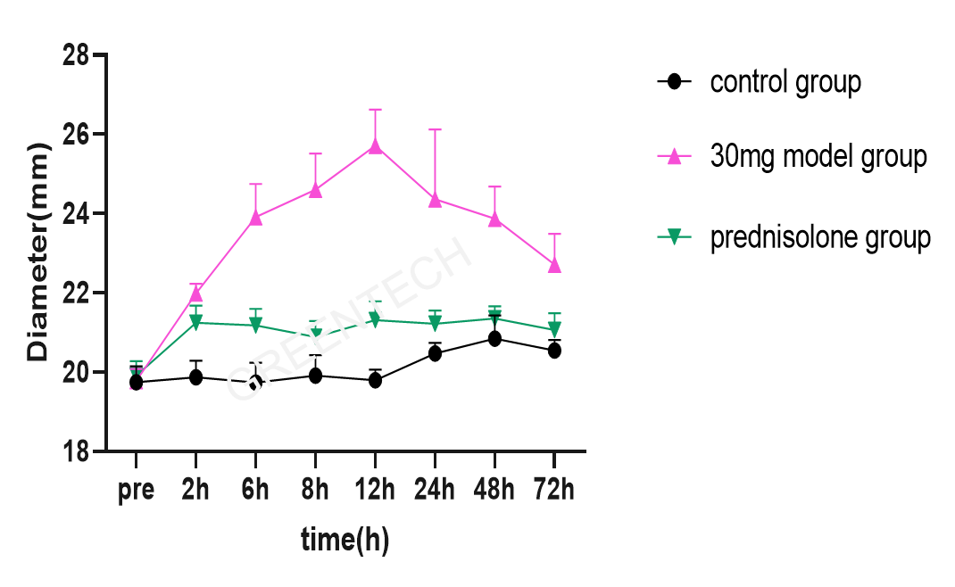
Figure 3. Evaluation of joint swelling in the rabbit model of gouty arthritis.

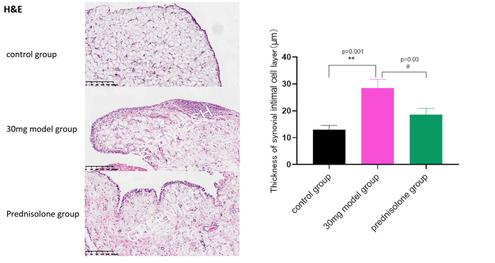
Figure 4. The thickness and histopathological assessment of synovial intima.
Inquiries
Request a quote now, or email us at BD@greentech-bio.com to inquire about our services or obtain a quote for your project.
Reference
Goo B, Lee J, Park C, Yune T, Park Y. Bee Venom Alleviated Edema and Pain in Monosodium Urate Crystals-Induced Gouty Arthritis in Rat by Inhibiting Inflammation. Toxins (Basel). 2021;13(9):661. Published 2021 Sep 16. doi:10.3390/toxins13090661


