Model of Multiple Sclerosis (MS)
Multiple sclerosis (MS) is a chronic demyelinating disease of the central nervous system (CNS) characterized by inflammatory infiltrates, demyelinating plaques, and axonal damage. Greentech Bioscience offers experimental autoimmune encephalomyelitis (EAE) model for mechanism studies and preclinical pharmacological studies of therapies against multiple sclerosis.
Experimental Autoimmune Encephalomyelitis (EAE) Model
Proteolipid protein (PLP) is an essential component for the formation and maintenance of the CNS myelin. Immunization of SJL/J mice with PLP139-151 peptide induces experimental autoimmune encephalomyelitis, an animal model of multiple sclerosis. The EAE mouse model has a complex neuropathology, and as with multiple sclerosis in humans, the condition in rodents exhibits a relapsing and remitting pattern and are characterized by nerve conduction defects and progressive disability. The EAE model is the classical animal model of MS, and has been widely used in the development, testing or validation of many drugs.
Animals: 8~10 week-old female SJL/J mice
Induction: Animals are immunized with PLP139-151 peptide emulsified in Complete Freund’s Adjuvant (CFA). Mice are injected intraperitoneally with pertussis toxin (PTX) immediately and two days after PLP139-151 immunization.
Clinical Assessment
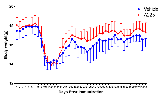
(1) Body weight
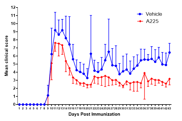
(2) Neurological function score (Weaver’s 15-point scoring system)
Case Study
EAE mouse model of multiple sclerosis

Figure 1. Body weight changes in an EAE mouse model.

Figure 2. EAE clinical scores.

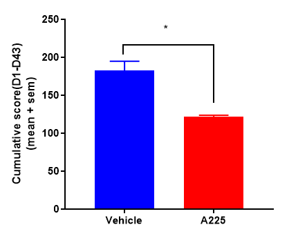
Figure 3. Cumulative scores of EAE models (vs Model group: *, P<0.05; n=14)
References
1. Stolz L,Derouiche A,Devraj K, et al. Anticoagulation with warfarin and rivaroxaban ameliorates experimental autoimmune encephalomyelitis.[J].J Neuroinflammation,2017,1:152.
2. Cruz-Herranz A,Dietrich M,Hilla AM, et al. Monitoring retinal changes with optical coherence tomography predicts neuronal loss in experimental autoimmune encephalomyelitis.[J].J Neuroinflammation,2019,1:203.
Inquiries
Request a quote now, or email us at BD@greentech-bio.com to inquire about our services or obtain a quote for your project.


