Models of Myocardial Hypertrophy and Heart Failure
Greentech Bioscience offers a comprehensive portfolio of animal models of hypertrophic cardiomyopathy and heart failure, from rodents to pigs, dogs, and non-human primates (NHPs). We also customize animal models to test the effectiveness of drugs according to our clients’ requirements.
Our Models of Cardiac Hypertrophy and Heart Failure
1. Animal Models of Ischemic Heart Failure
Models | Coronary Artery Occlusion (DSA technique) | Left Anterior Descending (LAD) Coronary Artery Ligation |
Species | Pigs/dogs/NHPs | Rats/pigs/dogs/NHPs |
Induction | Introduction of embolic agents into coronary artery based on DSA | LAD ligation |
Disease Characteristics | Irreversible symptoms such as left ventricular dysfunction, decreased ejection fraction, and neuro-endocrine activation | Effective replication of human acute and compensated heart failure after myocardial infarction |
Applications | Applied to research of myocardial ischemia and left ventricular remodeling after myocardial infarction | Applied to investigation of the cellular and subcellular mechanisms underlying the transition from myocardial infarction and myocardial hypertrophy to heart failure |
2. Pressure Overload-Induced Models of Heart Failure
Induction: transverse aortic constriction (TAC) and abdominal aortic constriction (AAC)
Species: rats, mice
Disease characteristics: increased aortic pressure and left ventricular pressure overload, replicating the pathological process from cardiac hypertrophy (about 4 weeks) to heart failure (about 12 weeks) caused by increased afterload
Applications: investigation of pathological changes and molecular mechanisms underlying heart failure caused by diastolic dysfunction and left ventricular hypertrophy
3. Drug-Induced Models of Heart Failure
Induction: tail vein injection of adriamycin
Species: rats
Disease characteristics: focal cardiomyocyte degeneration, edema, and vacuolation, as well as myocardial interstitial fibrosis, suggesting left ventricular remodeling and myocardial fibrosis
Applications: study of the pathogenesis of cardiomyopathy and heart failure and evaluation of novel treatments
4. Tachypacing-Induced Heart Failure Model
Induction: rapid right ventricular pacing (RRVP)
Species: dogs
Disease characteristics: progressive dilation of the ventricles, accompanied by diastolic dysfunction, significant decrease in ejection fraction and cardiac output, and increased peripheral vascular resistance
Applications: rapid ventricular pacing is an effective method to study the pathophysiological changes, molecular and biological characteristics, neuroendocrine changes and drug intervention during different stages of chronic heart failure
5. Volume Overload-Induced Models for Heart Failure
Induction: (thoracotomy) mitral regurgitation, arteriovenous fistula, inferior vena cava stenosis; (minimally invasive surgery) mitral insufficiency, aortic incompetence
Species: pigs, dogs, non-human primates (NHPs)
Clinical Assessment
- Ultrasonic cardiogram: left ventricular end-diastolic diameter (LVEDD), left ventricular end-systolic diameter (LVESD), left ventricular end-diastolic volume (LVEDV), left ventricular end-systolic volume (LVESV), left ventricular posterior wall thickness (LPwT), ejection fraction (EF), cardiac output (CO), FS, SV, chamber dimensions and wall thickness, pulmonary artery pressure, etc.
- Invasive hemodynamic monitoring: central venous pressure (CVP), right atrial pressure (RAP), right ventricular pressure (RVP), pulmonary artery pressure (PAP), Pulmonary capillary wedge pressure (PCWP), cardiac output (CO)
- 12-lead electrocardiogram
- Myocardial enzymogram and biochemical examination
- Histopathology
Basic Service
Body weight, food intake, excretion
Blood and tissue collection
Optional Endpoint
Imaging examinations such as MRI
Coronary angiography (for large animals)
Case Study
Figure 1. Echocardiographic measurements in SD rats before and one week after LAD ligation.

Figure 2. Cardiac function parameters in SD rats after LAD ligation.

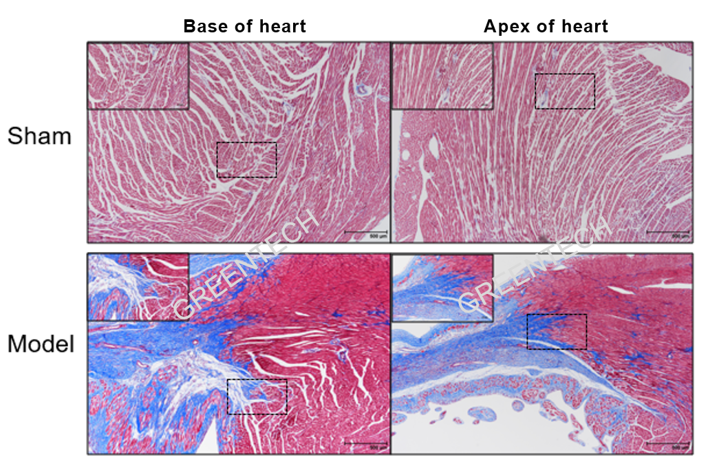
Figure 3. LAD ligation-induced myocardial infarction rats (Masson staining).

Figure 4. Cardiac function parameters in rhesus monkeys after LAD ligation.
Inquiries
Request a quote now, or email us at BD@greentech-bio.com to inquire about our services or obtain a quote for your project.
References
1. Conceição, G., Heinonen, I., Lourenço, A. P., Duncker, D. J., & Falcão-Pires, I. (2016). Animal models of heart failure with preserved ejection fraction. Netherlands Heart Journal, 24(4), 275–286. doi:10.1007/s12471-016-0815-9
2. Janssen, P. M. L., & Elnakish, M. T. (2019). Modeling heart failure in animal models for novel drug discovery and development. Expert Opinion on Drug Discovery, 1–9.


